Dental implants are an excellent long-term option to restore the esthetics and function of your smile. In fact, their development and use are among the greatest advances in dentistry over the last 40 years.
Call or message us to schedule an appointment!
A dental implant is a very effective and safe alternative for replacing missing teeth because it functions like an artificial root inserted into the bone so it both looks and feels like a natural tooth.
Once the implant is placed in the bone, a natural osseointegration process begins and lasts a few months. When this process is complete, an abutment is connected to the implant to support the dental crown.
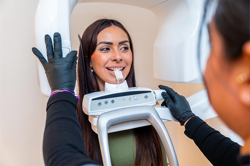
To determine whether a patient is a candidate for dental implants, it is essential that the periodontist review the results of a dental Cone Beam scan.
What is a Dental Cone Beam study?
Cone Beam Computed Tomography (CBCT) is a special type of X-ray that, in a single exposure, produces 3D images of the teeth, soft tissues, bones, and nerves.
Cone Beam is requested for all patients who need or want dental implants. The study is fundamental for the periodontist to establish a treatment plan and a surgical guide, as well as to determine whether there is sufficient bone and space to place implants successfully.
Cone Beam has revolutionized imaging in medicine, oral pathology, and oral surgery.
Differences between Cone Beam and a dental TAC
Although a 3D dental CT can yield similar results, there are important differences between these two radiologic exams.
- The first and likely most important difference is the radiation dose: with Cone Beam, it is much lower than with a conventional dental CT.
- Cone Beam produces a 3D image via a computerized process connected to the scanner. Traditional dental CT reconstructed a 3D image from a gradual rotation of the patient’s head, compiling multiple slices as the head turned.
- Cone Beam provides projections from all angles, offering a holistic image of the patient with just one 360-degree rotation.
- The images are isotropic (same properties in all directions), so they are free of distortion and notably clear and precise thanks to a direct CdTe (cadmium telluride) sensor.
- Exposure time to X-rays is also shorter with Cone Beam typically under one to two minutes, depending on the machine making it the better option when balancing image quality, time, and dose.
- Both tests emit X-rays; however, Cone Beam uses a conical beam, while the dental CT uses a spiral beam pattern.
- Lastly, while costs vary by imaging center, the dental CT (TAC) is generally more expensive than CBCT.
Other uses of the Cone Beam study
Beyond determining exact implant positioning, bone structure, and tooth orientation, CBCT is also very useful to:
- Plan and manage orthodontic cases.
- Locate the source of oral or facial pain.
- Plan surgeries.
- Diagnose temporomandibular joint (TMJ) disorders.
- Evaluate the jaws, paranasal sinuses, neurovascular canals, and nasal cavity.
- Detect, measure, and assist in treating jaw tumors.
- Perform cephalometric analysis and orthognathic surgery planning.
Where can I get a CBCT?
CBCT scans are readily available at most radiology centers. Pricing typically ranges from MXN $1,500 to $3,000, and results are generally considered valid for six months.
If you’re interested in dental implants but have questions about cost, treatment time, or surgical requirements, call or message us and we’ll be happy to help.
This article explains the role of Cone Beam (CBCT) in modern dentistry, especially for implant planning. As noted, this study is essential for determining whether you have enough bone and for designing the surgery. This content is informational; the next step is a consultation to review your results and define a treatment plan.
Start your journey toward a safe, well-planned implant treatment. Request a consultation with Dr. Daniel Fernández via WhatsApp, phone, or our contact form.